


 301 / 536

2.2 Layout rules for nuclear medicine
facilities
Given the radiation protection constraints involved
in the use of unsealed radioactive sources, nuclear
medicine units are designed and organised so that
they can receive, store, prepare and then administer
unsealed radioactive sources to patients or handle
them in laboratories (radioimmunology for instance).
Provision is also made for the collection, storage and
disposal of radioactive wastes and effluents produced
in the facility, particularly the radionuclides contained
in patients’ urine.
From the radiological viewpoint, the personnel are
subjected to a risk of external exposure, in particular
on the fingers, due in particular to the handling of
certain radionuclides (case with fluorine-18, iodine-131
or yttrium-90), and a risk of internal exposure through
accidental intake of radioactive substances. Given these
conditions, the nuclear medicine units must satisfy the
rules prescribed by ASN resolution 2014-DC-0463 of
23rd October 2014 relative to the minimum technical
rules of design, operation and maintenance that
in vivo
nuclear medicine facilities must satisfy, approved by
the Order of 16th January 2015.
This resolution more specifically introduces new rules
for the ventilation of nuclear medicine units (cancellation
of the negative pressure requirements and hourly air
renewal rates figuring in the Order of 30th October
1981) and of the rooms accommodating patients being
treated for thyroid cancer with iodine-131 in particular
(new negative pressure requirement). Furthermore, the
facilities equipped with a CT scanner coupled with a
gamma camera or a PET camera must comply with the
provisions of ASN resolution 2013-DC-0349 of 4th June
2013 (see chapter 3). This resolution requires that the
layout and access to these facilities comply with the
radiation protection rules set by French Standard NFC
15-160 in its March 2011 version
3. EXTERNAL-BEAM RADIOTHERAPY
AND BRACHYTHERAPY
3.1 Description of the techniques
Alongside surgery and chemotherapy, radiotherapy is
one of the key techniques employed to treat cancerous
tumours. Some 175,000 patients are treated each year.
Radiotherapy uses ionising radiation to destroymalignant
cells (and non-malignant cells in a small number of
cases). The ionising radiation necessary for treatment is
either produced by an electric generator or emitted by
radionuclides in the form of a sealed source. There are
thus two ways of delivering the radiation: external-beam
radiotherapy, where the source of radiation produced
by a particle accelerator or radioactive sources (Gamma
knife
®
for example) is external to the patient, and
brachytherapy, where the source is placed in direct
contact with the patient, within or as close as possible
to the area to treat.
At the end of 2014, external beam radiotherapy
installations comprised 476 treatment devices, including
461 conventional linear accelerators. These devices are
installed in 176 radiotherapy centres, of which roughly
half have public status and the other half private status.
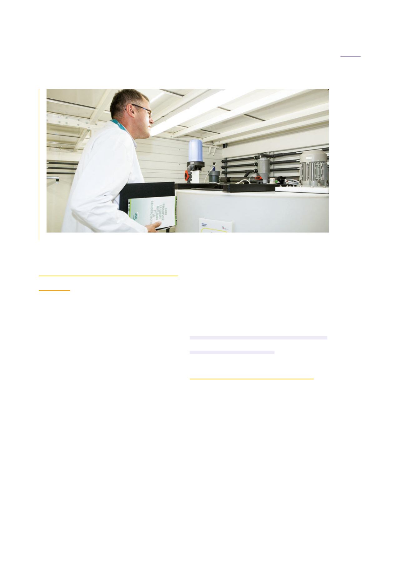
ASN inspection of the nuclear medicine unit of the Eugène-Marquis regional cancer centre in Rennes, July 2015.
301
CHAPTER 09:
MEDICAL USES OF IONISING RADIATION
ASN report on the state of nuclear safety and radiation protection in France in 2015