


 302 / 536

There are six hundred and fifty three (653) registered
radiation oncologists (source: French Radiotherapy
Observatory).
3.1.1 External-beam radiotherapy
The irradiation sessions are always precededby preparation
of a treatment planwhich defines the dose to be delivered,
the target volume(s) to be treated, the irradiation beam
setting and the estimated dose distribution (dosimetry) for
each patient. Preparation of this plan, which aims to set
conditions for achieving a high dose in the target volume
while preserving surrounding healthy tissues, requires
close cooperation between the radiation oncologist, the
medical physicist and, when applicable, the dosimetrists.
In the vast majority of treatments, irradiation is ensured
using linear particle accelerators with an isocentric arm
emitting beams of photons produced at a voltage varying
from4 to 25megavolts (MV) or electrons with an energy
level of between 4 and 25megaelectronvolts (MeV) and
delivering dose-rates that can vary from 2 to 6 grays per
minute (Gy)/min, although some latest-generation linear
accelerators can deliver much higher dose-rates of up to
25 Gy/min (in the case of photon beams).
For certain specific therapeutic indications, several centres
propose treatments that are made possible thanks in
particular to the use of:
•
a linear accelerator equipped with specific functions
(micromultileaf collimator, additional imaging systems,
robotic arm and/or table, etc.);
•
a gammatherapy device equipped with more than
200 sources of cobalt-60;
•
a cyclotron producing proton beams.
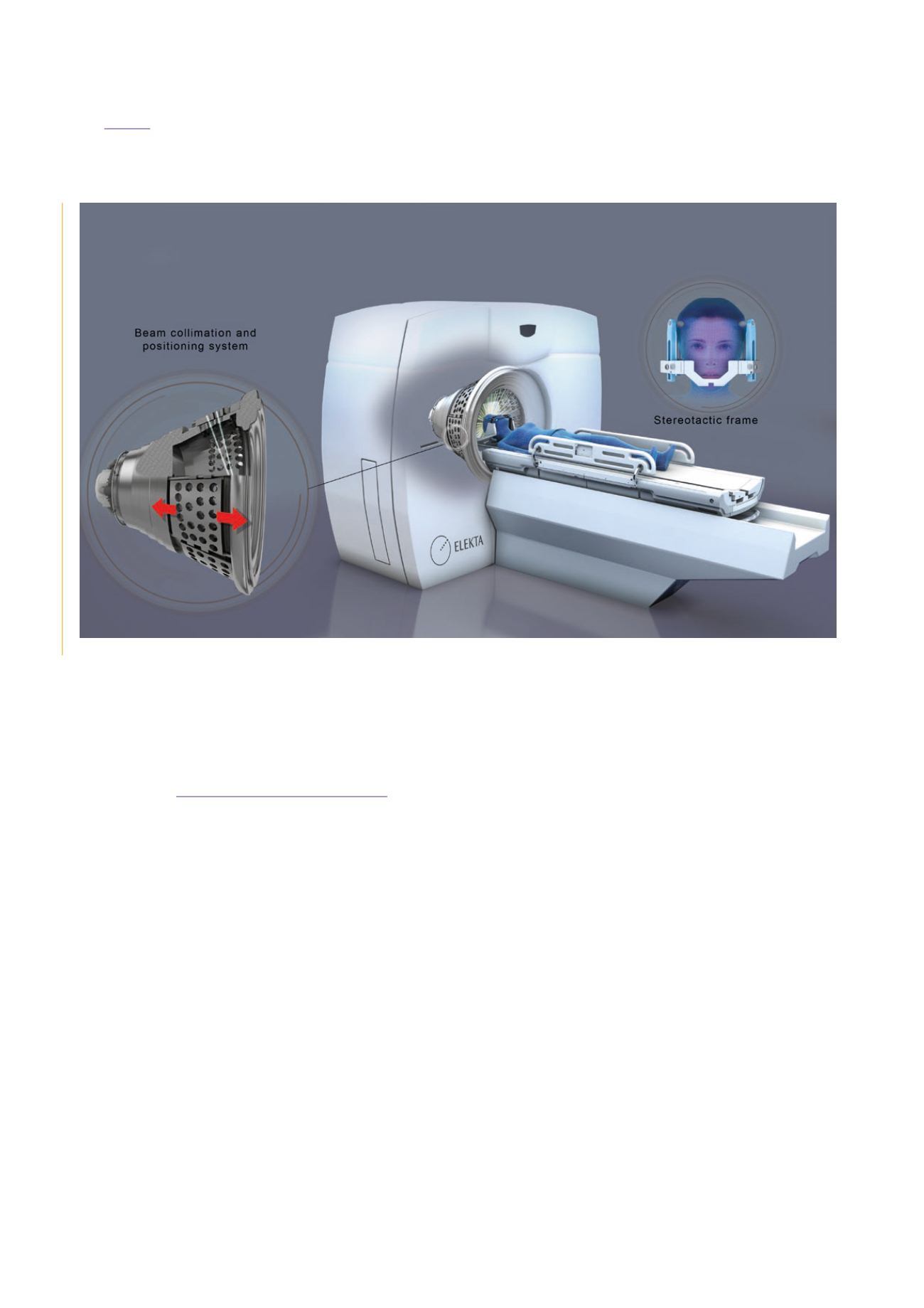
Stereotactic radiotherapy
Stereotactic radiotherapy is a treatment method which
aims to offer millimetre-precise, high-dose irradiation
using multiple mini-beams converging in the centre of
the target, for intra- or extra-cranial lesions. In stereotactic
radiotherapy treatments, the total dose is delivered either in
a single sessionor in a hypofractionatedmanner, depending
on the disease being treated. The termradiosurgery is used
to designate treatments carried out in a single session.
This technique firstly requires great precision in defining
the target volume to irradiate, and secondly that the
treatment be as conformal as possible, that is to say that
the irradiation beams follow the shape of the tumour as
closely as possible.
It was originally developed to treat surgically-inaccessible
non-cancerous diseases in neurosurgery (artery or vein
malformations, benign tumours) and uses specific
positioning techniques to ensure very precise localisation
of the lesion.
Elekta Gamma Knife® system used in intracranial radiosurgery and radiotherapy.
302
CHAPTER 09:
MEDICAL USES OF IONISING RADIATION
ASN report on the state of nuclear safety and radiation protection in France in 2015